A Study of Toxicity Evaluation Using the iMScope TRIO - Analysis of Localization of Amiodarone in Rat Lungs -
In recent years, the analytical technique of MS imaging has been attracting attention as a method that can detect data on the localization of candidate compounds without using a label.
This method is expected to provide a breakthrough in drug discovery research as it can be used to analyze the localization of various substances without labeling, and to simultaneously analyze the unchanged drug and its metabolites using the same section.
Here, we introduce an example of MS imaging analysis using the iMScope TRIO imaging mass microscope to compare the localization of the pathological findings with that of the amiodarone observed in lung tissue after administering amiodarone (Fig. 1) to rats.
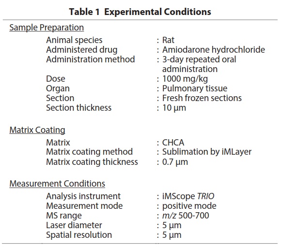
In this experiment, we measured the tissue sections of rats lungs that had been administered amiodarone, an anti- arrhythmia drug. When administered in large quantities, amiodarone causes phospholipidosis and pathological findings such as foamy macrophage infiltration of cells are observed. However, up until now there had been no information on whether amiodarone accumulated in the lesions or not, so we examined the relationship between the pathological findings and localization of amiodarone utilizing the MS imaging technique. In the preliminary study test using standard amiodarone, we optimized the matrix selection and measurement mode, and applied those conditions in this experiment. Table 1 shows the experimental conditions from sample preparation to MS imaging analysis.
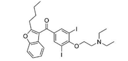
Fig.1 Structural Formula of Amiodarone
iMScope QT
Features:
The instrument is a combination of an optical microscope which allows the observation of high-resolution morphological images, with a mass spectrometer which identifies and visualizes the distribution of specific molecules.
Superimposing the two images obtained based on these very different principles,has created a significant new research tool, the imaging mass microscope.